Gadgets
3D scans reveal secrets of a 3,000-year-old Egyptian mummy’s coffin

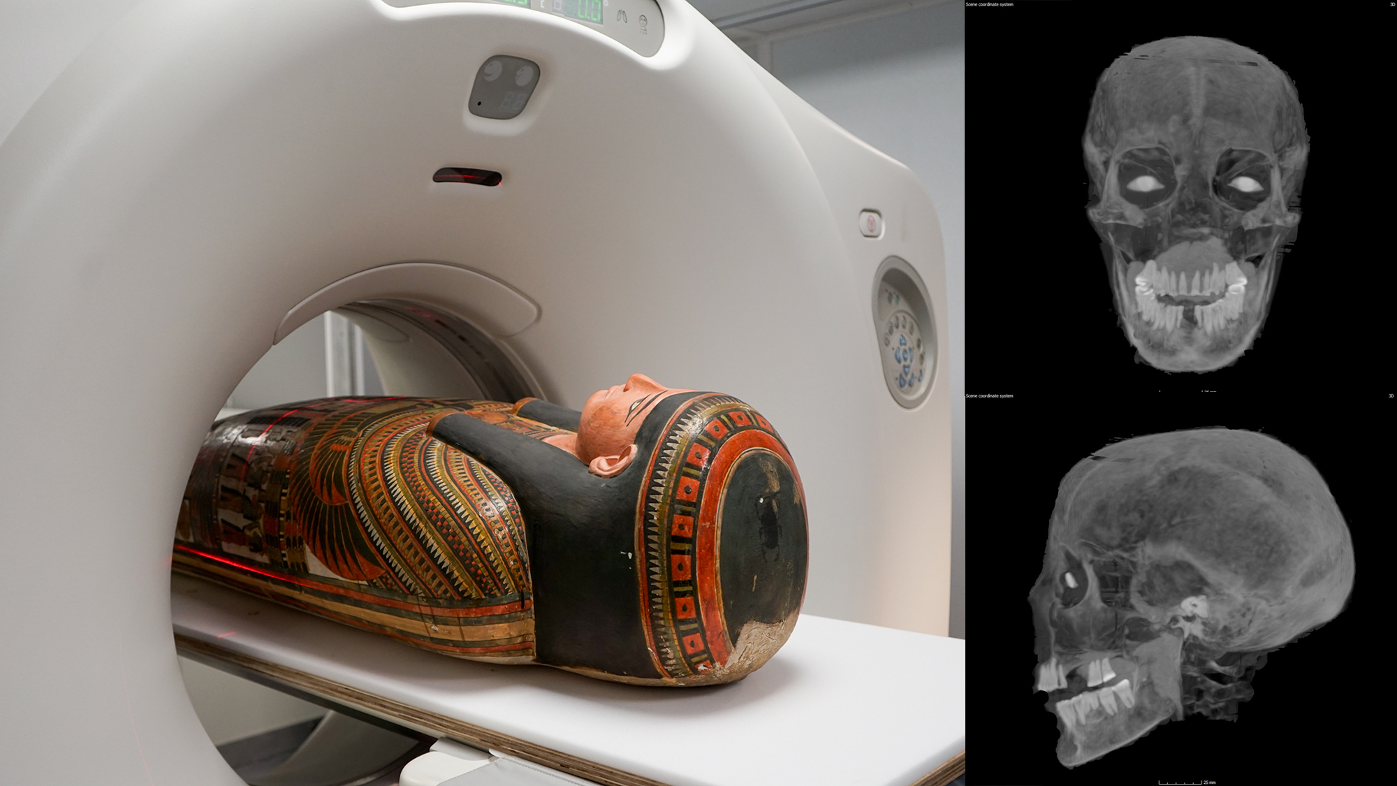
Researchers at Chicago’s Field Museum have long been intrigued by the burial procedure of Lady Chenet-aa, one of the museum’s ancient Egyptian mummies. Recently, the mystery surrounding her burial has been unraveled using a CT scanner.
Lady Chenet-aa lived during Egypt’s Third Intermediate Period approximately 3,000 years ago. Following her death, she was prepared for the afterlife by being encased in a cartonnage, a type of paper mache box commonly used for mummies. However, what puzzled experts was the seamless appearance of her casing, without any visible seams. Thanks to a recent announcement from the Field Museum on October 24, a mobile CT scanner has shed light on how Chenet-aa was placed inside the “locked-mummy” cartonnage.

CT scanning, which generates 3D images by stacking X-ray scans digitally, provided detailed insights into Chenet-aa’s cartonnage and burial. By transporting 26 mummies, including Chenet-aa, to a mobile CT scanner outside the museum, experts were able to analyze the casing and its contents with unprecedented clarity. This revealed the intricate details of how Chenet-aa was placed inside the seemingly seamless cartonnage.
“You can start to see that there’s a seam going down the back and some lacing,” stated JP Brown, the museum’s senior conservator of anthropology.

The CT scans also provided new insights into Chenet-aa’s health at the time of her death. Researchers estimate she passed away in her late 30s or early 40s, although the exact cause of death remains unknown. Her teeth showed signs of wear and damage, with many missing and the remaining teeth exhibiting significant wear. This dental condition is attributed to the presence of sand particles in her food, which eroded the enamel over time.

CT scans also revealed artificial eyes placed in Chenet-aa’s eye sockets, despite the absence of natural eyes. These eyes, made of an unknown material, symbolize the belief in providing the deceased with sight in the afterlife.
[Related: ‘Screaming woman’ may solve a 3,500-year-old mummy mystery.]
“The ancient Egyptian view of the afterlife is similar to our ideas about retirement savings. It’s something you prepare for, put money aside for all the way through your life, and hope you’ve got enough at the end to really enjoy yourself,” Brown explained. “The additions are very literal. If you want eyes, then there needs to be physical eyes, or at least some physical allusion to eyes.”
These findings mark the beginning of a deeper exploration into the secrets of the Field Museum’s ancient mummies. Over the coming year, researchers plan to delve further into the wealth of CT scans for additional revelations about life and death in ancient Egypt.
Please rewrite the text for me.
-

 Destination8 months ago
Singapore Airlines CEO set to join board of Air India, BA News, BA
-

 Breaking News10 months ago
Croatia to reintroduce compulsory military draft as regional tensions soar
-

 Gadgets3 months ago
Supernatural Season 16 Revival News, Cast, Plot and Release Date
-

 Tech News12 months ago
Bangladeshi police agents accused of selling citizens’ personal information on Telegram
-

 Productivity11 months ago
How Your Contact Center Can Become A Customer Engagement Center
-

 Gadgets4 weeks ago
Fallout Season 2 Potential Release Date, Cast, Plot and News
-

 Breaking News10 months ago
Bangladesh crisis: Refaat Ahmed sworn in as Bangladesh’s new chief justice
-

 Toys12 months ago
15 of the Best Trike & Tricycles Mums Recommend


